Topcon Fundus Camera NW8
Price range: $2,925.00 through $2,950.00
Code: TRC-NW8
Brand:Topcon
Product Condition: Refurbished
Description
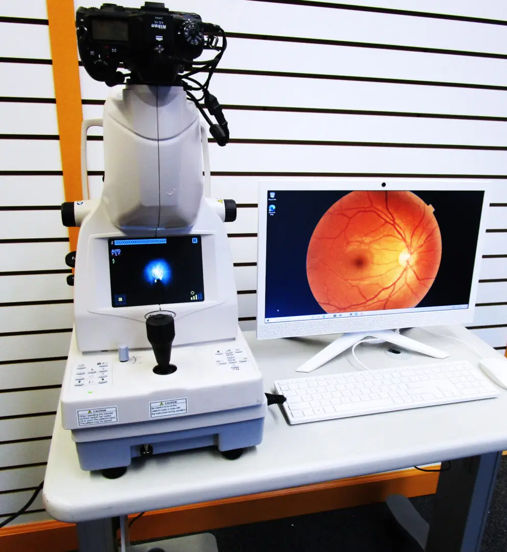
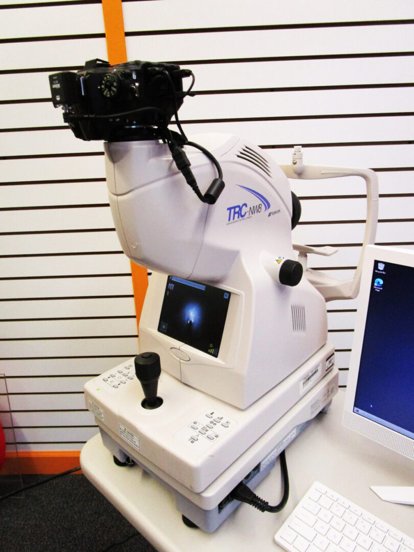
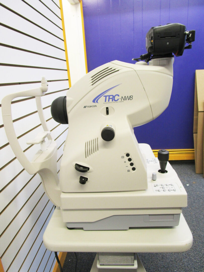
Topcon Fundus Camera NW8
Includes:
- Topcon Fundus Camera NW8
- Nikon Top Camera
- HP All-In-One Computer with 21.5 In Screen
- Topcon Easy Capture Software
- Interconnect Cables
- Power Cords
Image on Website is Actual Unit Image
Topcon Fundus Camera – NW8 Non-Mydriatic Fundus Camera The Topcon TRC-NW8 is an easy-to-use, auto focus, auto capture non-mydriatic imaging system that is designed to obtain high resolution color and monochrome images of the retina and the anterior segment of the human eye. It is equipped with automatic focusing and capture making its use quick and simple. A 12.3 megapixel camera back provides high resolution images with a 45° field of view. The incorporated filters and internal firmware allow the user to obtain color, red-free and fluorescein angiography images. The TRC-NW8F has nine internal fixation points that facilitate the composition of wide angle views of the retina. The timer feature allows the user to add a time stamp on each captured image.
Retina photography, also known as fundus photography, represents a significant advancement in ophthalmic imaging technology, providing detailed and non-invasive visualization of the retina. This essay explores the principles, applications, and benefits of retina photography in clinical practice and research.
Principles of Retina Photography and Fundus Cameras:
Retina photography involves capturing high-resolution images of the retina, which is the light-sensitive tissue lining the back of the eye. The key principles underlying retina photography include:
- Light Source and Optics: Fundus cameras utilize specialized optics and illumination systems to capture clear images of the retina. Typically, these cameras employ a combination of visible light or near-infrared light to illuminate the retina without causing discomfort to the patient.
- Digital Imaging Sensors: Modern retina cameras are equipped with high-resolution digital sensors that capture detailed images of the retina. These sensors ensure clarity and precision in imaging, allowing ophthalmologists to observe subtle changes in retinal structures.
- Field of View: Retina cameras offer different fields of view, ranging from small areas for detailed examination of the macula (central part of the retina) to wide-angle views for assessing peripheral retina health.
- Image Processing and Analysis: Images obtained through retina photography can be processed and analyzed using software tools. This aids in quantifying changes in retinal features, such as blood vessels, optic nerve, and retinal pigment epithelium, which are critical for diagnosing and monitoring various eye conditions.
Applications of Retina Photography:
Retina photography finds extensive application across various domains of ophthalmology and eye care:
- Diagnosis and Monitoring of Eye Diseases: Retina photography is invaluable in diagnosing and monitoring conditions such as diabetic retinopathy, age-related macular degeneration (AMD), glaucoma, retinal detachments, and hypertensive retinopathy. These images serve as baseline references and enable ophthalmologists to track disease progression over time.
- Screening Programs: In public health initiatives, retina photography plays a crucial role in screening large populations for diabetic retinopathy and other sight-threatening conditions. Automated systems can analyze images to identify abnormalities, facilitating early intervention and reducing the risk of vision loss.
- Research and Education: Retina photography supports research efforts by providing visual documentation of eye diseases and treatment outcomes. It serves as an educational tool for training medical students, residents, and ophthalmic technicians in recognizing retinal pathologies and interpreting imaging findings.
- Telemedicine and Remote Consultations: With advancements in telemedicine, retina photography allows remote ophthalmologists to assess and provide consultations on retinal health. Images can be securely transmitted for expert review, enhancing access to specialized eye care in underserved regions.
Benefits of Fundus Photography and Fundus Camera:
The adoption of retina photography offers several benefits to patients, clinicians, and the healthcare system:
- Early Detection and Intervention: By detecting subtle changes in the retina early, retina photography enables prompt intervention and management of eye diseases, thereby preserving vision and preventing irreversible damage.
- Objective Documentation: Images captured through retina photography provide objective documentation of retinal findings, reducing reliance on subjective assessments and enhancing consistency in clinical evaluations.
- Patient Engagement and Education: Visual representation of retinal health fosters patient engagement by enabling individuals to better understand their condition and treatment options. It promotes informed decision-making and compliance with recommended eye care regimens.
- Efficiency and Cost-Effectiveness: Compared to traditional methods, retina photography is efficient, non-invasive, and relatively quick. It reduces the need for invasive procedures and frequent follow-up visits, thereby optimizing healthcare resources and reducing overall healthcare costs.
Conclusion:
In conclusion, retina photography stands at the forefront of diagnostic imaging technology in ophthalmology, revolutionizing the assessment and management of various eye diseases. By providing detailed, real-time visualization of the retina, it empowers clinicians to deliver timely interventions and personalized care to patients. As technology continues to advance, retina photography holds promise for further enhancing diagnostic accuracy, expanding access to eye care services, and improving outcomes for individuals worldwide.
See Article:
Additional information
| Weight | 45 lbs |
|---|---|
| Dimensions | 65 × 24 in |
| Computer Type | Desktop All-In One, Laptop |
Topcon NW8 Information
Topcon NW8 Image

The Retina
You must be logged in to post a review.
Related products
-
Sale!

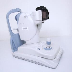
Canon CR-2 Digital Retinal Camera
Original price was: $1,950.00.$1,250.00Current price is: $1,250.00. Read more -
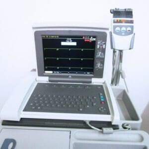
GE Mac 5500HD ECG with Cam, Leads and Wifi
$1,499.00 Add to cart -
Welch Allyn Wall Mounted Otoscope 777 Wall Transformer
Price range: $299.00 through $499.00 Select options This product has multiple variants. The options may be chosen on the product page -
Sale!
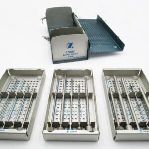
CORB Biopsy Needle Set Ref. 3240-62
Original price was: $129.00.$99.00Current price is: $99.00. Add to cart

Reviews
There are no reviews yet.